


What Is Ultrasonography and How Does It Work?
Ultrasonography, also known as ultrasound imaging, is a widely used diagnostic technique in modern medicine. It employs high-frequency sound waves to produce real-time images of internal organs, tissues, and blood flow. This non-invasive, painless, and relatively inexpensive method plays a critical role in detecting and diagnosing a wide variety of medical conditions. From pregnancy monitoring to diagnosing heart and abdominal issues, ultrasonography has revolutionized the way healthcare professionals examine the human body.
Understanding the Basics of Ultrasonography
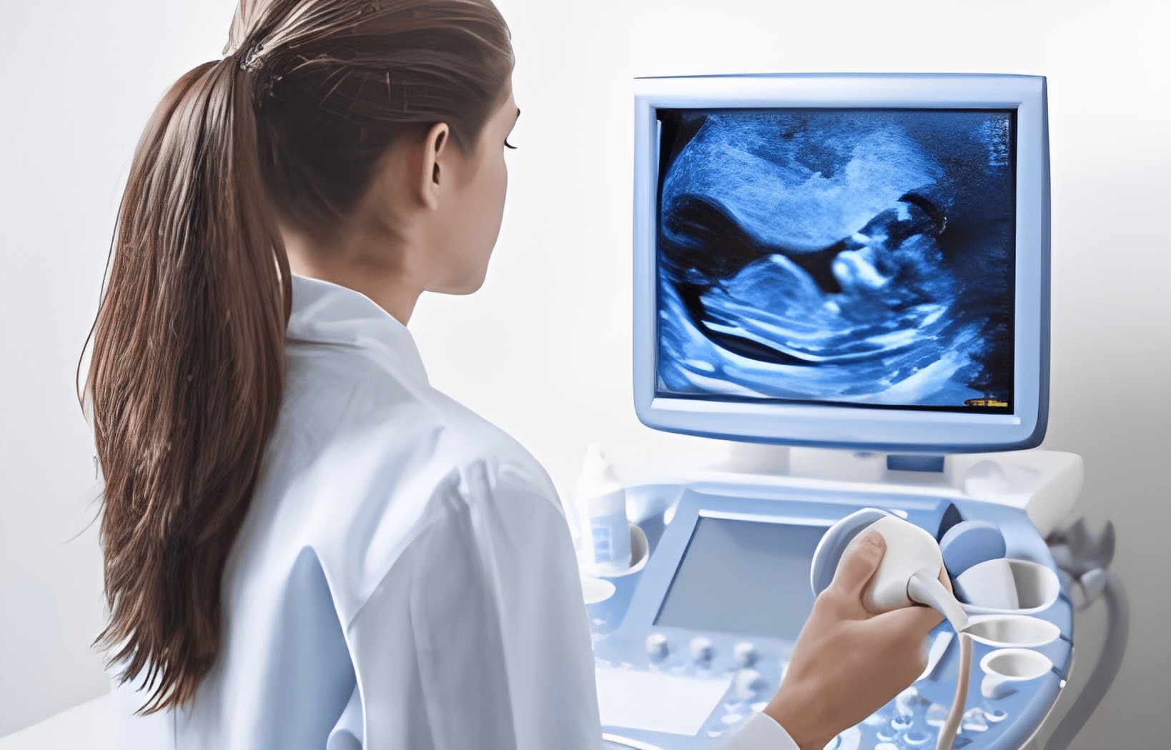
Ultrasonography operates based on the principles of sound wave reflection. A device called a transducer emits high-frequency sound waves that travel through the body. When these sound waves encounter tissues, fluids, or organs, they bounce back to the transducer. The returning echoes are then converted into visual images by a computer. These images are displayed in real-time on a monitor, allowing healthcare providers to observe movement, such as a beating heart or flowing blood.
The History and Evolution of Ultrasonography
Ultrasonography originated in the mid-20th century, inspired by sonar technology developed during World War II. Initially used primarily in obstetrics to visualize the developing fetus, it gradually expanded into many other areas of medicine. Advances in digital technology have significantly improved the clarity and resolution of ultrasound images. Today, ultrasonography is used not only for diagnostic purposes but also for guiding interventional procedures such as biopsies and needle aspirations.
Key Components of Ultrasonography Equipment
The Transducer
The transducer is the handheld device that emits and receives sound waves. It contains piezoelectric crystals that generate ultrasound waves when electrified. These same crystals detect the returning echoes and convert them into electrical signals.
The Central Processing Unit (CPU)
The CPU is the brain of the ultrasound machine. It processes the signals received from the transducer, converting them into images through complex mathematical calculations known as digital signal processing.
The Display Monitor
The monitor displays the real-time images that are generated. Modern ultrasound machines offer high-definition screens and color Doppler capabilities to visualize blood flow and enhance diagnostic precision.
Data Storage
Ultrasound systems often include internal or external storage options to save images and videos. These records are essential for tracking changes over time and supporting medical documentation.
Different Types of Ultrasonography
Abdominal Ultrasonography
This type is used to visualize organs within the abdominal cavity, including the liver, gallbladder, pancreas, kidneys, and spleen. It is commonly used to detect gallstones, liver disease, and kidney stones.
Obstetric and Gynecologic Ultrasonography
Used extensively during pregnancy, obstetric ultrasonography monitors fetal development, estimates gestational age, and evaluates the health of the placenta and amniotic fluid. Gynecologic ultrasound helps assess uterine and ovarian conditions.
Cardiac Ultrasonography (Echocardiography)
Echocardiography evaluates the structure and function of the heart. It helps diagnose heart valve issues, cardiac output problems, and congenital heart defects.
Musculoskeletal Ultrasonography
This type is used to examine muscles, tendons, and ligaments. It is commonly used in sports medicine to assess injuries and inflammation.
Vascular Ultrasonography
It visualizes blood vessels and helps identify conditions such as blood clots, blocked arteries, and varicose veins using Doppler techniques to assess blood flow.
Pelvic Ultrasonography
This procedure is performed to examine the bladder, prostate, uterus, and ovaries. It is often used to diagnose urinary tract issues, reproductive organ abnormalities, or pelvic pain.
How Ultrasonography Is Performed
The procedure typically begins with the patient lying on an examination table. A water-based gel is applied to the skin over the area to be examined. This gel eliminates air between the skin and transducer to ensure sound waves penetrate the body efficiently. The transducer is then moved gently over the skin, sending sound waves and receiving echoes. The entire procedure usually takes between 15 to 45 minutes, depending on the complexity of the examination.
Advantages of Ultrasonography
Non-Invasive and Safe
Unlike X-rays and CT scans, ultrasonography does not use ionizing radiation, making it safe for repeated use and suitable for pregnant women and children.
Real-Time Imaging
It provides dynamic imaging, allowing observation of movement, which is especially beneficial for cardiac and fetal assessments.
Portable and Accessible
Portable ultrasound machines can be used in emergency rooms, ambulances, and even in remote locations, making ultrasonography highly accessible.
Cost-Effective
Compared to other imaging modalities such as MRI or CT, ultrasonography is relatively low-cost, making it ideal for widespread use in medical facilities.
Limitations of Ultrasonography
Limited Penetration
Ultrasonography is less effective in imaging structures located behind bones or gas-filled organs such as the lungs or intestines.
Operator Dependency
The accuracy of ultrasonography greatly depends on the skill and experience of the operator. Inexperienced users may miss important findings or produce unclear images.
Lower Image Resolution
Although image quality has improved significantly, it still does not match the resolution provided by CT or MRI, particularly for deep tissue visualization.
Innovations and Future Trends in Ultrasonography
With the advent of artificial intelligence and machine learning, the future of ultrasonography is promising. AI algorithms can assist in image interpretation, reducing human error and improving diagnostic accuracy. Additionally, advances in 3D and 4D ultrasonography provide more detailed anatomical views and real-time movement, which are particularly useful in fetal and cardiac imaging.
Portable and handheld ultrasound devices are becoming more powerful and affordable, democratizing access to ultrasonography even in low-resource settings. Telemedicine integration allows remote interpretation of ultrasound images, expanding its reach in rural and underserved areas.
Conclusion
Ultrasonography has become an indispensable tool in modern medical diagnostics. Its non-invasive nature, safety profile, and real-time imaging capabilities make it ideal for a wide range of applications across multiple specialties. While it has certain limitations, ongoing technological advancements continue to enhance its accuracy and usability. Understanding how ultrasonography works not only deepens our appreciation of medical technology but also empowers patients to make informed decisions about their health care. Whether monitoring a pregnancy or diagnosing a complex condition, ultrasonography remains at the forefront of medical imaging.